Home » Without Label » Tendon Diagram - Knee Anatomy : On the other hand, the insertion is where a tendon attaches that muscle to the *more* movable bone.
Tendon Diagram - Knee Anatomy : On the other hand, the insertion is where a tendon attaches that muscle to the *more* movable bone.
Tendon Diagram - Knee Anatomy : On the other hand, the insertion is where a tendon attaches that muscle to the *more* movable bone.. On the other hand, the insertion is where a tendon attaches that muscle to the *more* movable bone. Possibly the most important tendon in terms of mobility is the achilles tendon. Fall on one point of shoulder and can rupture these ligaments with dislocation of ac joint. These structures work together to support the body, enable a range of movements, and send messages from the brain to. Flexor tendon lacerations are classified into five zones 2, 15, 16.
When the muscles tighten (contract) arguably, the most important tendon is the achilles tendon, which allows the calf muscles to move. The tendon is firmly connected to muscle fibres at one end and to components of the bone at its other end. Foot anatomy diagram, foot joint diagram, foot sprain diagram, foot tendons and ligaments pain, leg tendon diagram, peroneal tendonitis, foot, foot anatomy diagram, foot joint diagram, foot sprain diagram, foot tendons and ligaments pain, leg tendon diagram, peroneal tendonitis. Bones, cartilage, ligaments, and tendons. The tendon travels along the inside of the forearm on the side of the small finger and crosses the wrist.
Patellar Tendon Tear Orthoinfo Aaos from orthoinfo.aaos.org 9 photos of the foot tendons and ligaments diagram. The achilles tendon is a tough band of fibrous tissue that connects the calf muscles to the heel bone (calcaneus). The achilles tendon enables us to walk, without it we would not be able to raise our heels of the ground. For images of the muscle, click on each link under location. The coracobrachialis muscle lies deep to the biceps brachii in the arm. Raises heal when leg is straight. There are over two dozen gorgeous and painstakingly detailed illustrations on this chart, from the extensor pollicis longus to the flexor digitorum. The bones together make up the hip.
The tendon is firmly connected to muscle fibres at one end and to components of the bone at its other end.
Superficial posterior muscles of the forearm posterior compartment muscles of the forearm. It is controlled by the obturator nerve. You can see a diagram of the achilles tendon below. Foot anatomy diagram, foot joint diagram, foot sprain diagram, foot tendons and ligaments pain, leg tendon diagram. Jul 05, 2018 · the foot diagram has a complex structure made up of bones, ligaments, muscles, and tendons. Flexes elbow and moves forearm. The achilles tendon enables us to walk, without it we would not be able to raise our heels of the ground. Brings hip away from body. Also allows the action of raising up onto toes. The coracobrachialis muscle lies deep to the biceps brachii in the arm. Tendon, tissue that attaches a muscle to other body parts, usually bones.tendons are the connective tissues that transmit the mechanical force of muscle contraction to the bones; The achilles tendon is the strongest and largest tendon in the body. Medical labeled diagram closeup with muscle, transverse carpal ligament, median nerve, tendon sheath, flextor tendons and bones.
The achilles tendon is also called the calcaneal tendon. Tendons are remarkably strong, having one of the highest tensile strengths found among soft tissues. It attaches to the wrist bone, the pisiform, and as well as the 5th hand bone. It is controlled by the obturator nerve. Ligaments join the knee bones and provide stability to the knee:
Knee Wikipedia from upload.wikimedia.org Tendons are thick bands of tissue that connect muscles to bones. One peroneal tendon attaches to the outer part of the midfoot, while the other tendon runs under the foot and attaches near the inside of the arch. Ligaments and tendons are adapted in response to changes in mechanical stiffness. Lower back muscle diagram anatomy does degenerative disc disease affect the lower back muscle? 17 photos of the diagram of shoulder muscles and tendons. Browse 318 hand anatomy tendons stock photos and images available, or start a new search to explore more stock photos and images. The fcu tendon is one of two tendons that bend the wrist. Bones, cartilage, ligaments, and tendons.
Also allows the action of raising up onto toes.
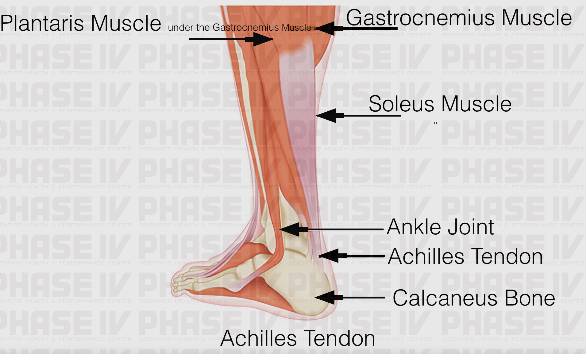
The changes in ligaments and tendons generally occur more slowly than adaptation in bone, because ligaments and tendons have less vascular supply. Extends spine and trunk back. The pubis, ischium, and ilium together constitute the pelvis while the thigh bone is the femur. The achilles tendon is a tough band of fibrous tissue that connects the calf muscles to the heel bone (calcaneus). When the muscles tighten (contract) arguably, the most important tendon is the achilles tendon, which allows the calf muscles to move. Tendon, tissue that attaches a muscle to other body parts, usually bones.tendons are the connective tissues that transmit the mechanical force of muscle contraction to the bones; Bones, cartilage, ligaments, and tendons. The bones together make up the hip. The two peroneal tendons in the foot run side by side behind the outer ankle bone. Tendon diagrams and design force vectors. Medical labeled diagram closeup with muscle, transverse carpal ligament, median nerve, tendon sheath, flextor tendons and bones. This important tendon in the back of the calf and ankle connects the plantaris, gastrocnemius, and soleus muscles to. The tendon is firmly connected to muscle fibres at one end and to components of the bone at its other end.
Tendons are thick bands of tissue that connect muscles to bones. Your biceps tendons attach the biceps muscle to bones in the shoulder and in the elbow. Extends spine and trunk back. Its muscle belly is in the forearm. Muscles and tendons of the human arm and hand, vintage engraved.
Tendon Anatomy Anatomy Drawing Diagram from phase-iv.com The achilles tendon enables us to walk, without it we would not be able to raise our heels of the ground. The tendon is firmly connected to muscle fibres at one end and to components of the bone at its other end. Tendons are found throughout the body, from the head and neck all the way down to the feet. Bones, cartilage, ligaments, and tendons. The ecu tendon works along with the ecrl and ecrb to straighten the wrist. Superficial posterior muscles of the forearm posterior compartment muscles of the forearm. These structures work together to support the body, enable a range of movements, and send messages from the brain to. Allows the foot to be turned inward and also supports the arch of the foot.
The ecu tendon works along with the ecrl and ecrb to straighten the wrist.
Your biceps tendons attach the biceps muscle to bones in the shoulder and in the elbow. Extends spine and trunk back. In the back and elsewhere in the body, tendons attach muscles to bones. If you tear the biceps tendon at the shoulder, you may lose some strength in your arm and have pain when you forcefully turn your arm from palm down to palm up. Intermediate back muscles and c. Ligaments join the knee bones and provide stability to the knee: Black and white print showing the musculoskeletal system of a human hand, including the bones, muscles, cartilage, tendons, ligaments, and joints,. The achilles tendon is also called the calcaneal tendon. Its muscle belly is in the forearm. The two peroneal tendons in the foot run side by side behind the outer ankle bone. The bones of the hip include the femur, the ilium, the ischium, and the pubis. The hand incorporates countless muscles, bones, tendons and ligaments into simple motion and this chart covers them all. Tendons are the connection between bones and muscles.